
North Queensland Knee
Experience Has No Substitute
Cartilage Repair
What is Articular Cartilage
Articular cartilage (AC) is highly specialised tissue on the ends of bones. Its function is to reduce friction and load. AC does not have a blood supply and gets all its nutrition from the joint fluid so its ability to repair itself is very limited. Arthritis is damage and loss of AC.
Cartilage Defect or Arthritis
A cartilage defect is an isolated divot or an area of damage in the cartilage of an otherwise normal cartilage surface. An isolated defect is an injury. Arthritis is a disease that involves opposing surfaces of a joint (this is called a kissing lesion). Despite much of what popular media would have you believe, arthritis is generally not amenable to repair procedures and is managed without surgery until the disease and associated symptoms are severe enough to warrant it.
If a cartilage defect is to be repaired multiple criteria must be satisfied for the repair to success. These include;
the size of the defect
the alignment of the limb (is the leg straight)
the stability of the joint (is the ACL intact)
the state of the meniscus (the cushion that will protect the repaired area).
Cartilage repair is therefore often combined with other procedures such as ACL reconstruction and osteotomy.
Cartilage Repair Procedures
A number of techniques are utilised to repair cartilage depending on the underlying problem. These techniques can be used to grow new cartilage (microdrilling, MACR, MACI) , repair existing cartilage back to the underlying bone (osteochondral repair) or transplant cartilage and bone into a defect (autologous osteochondral transfer).
Microfracture / Microdrilling
Microfracture is a minimally invasive procedure that was developed in the 1980's. It utilises your own stem cells in the bone beneath the cartilage to make repair cartilage (yes, stem cells are nothing new in orthopaedics - we've been harnessing their potential to repair the body for 40 years). A planktonic stem cell (one floating around in the joint) will not make cartilage but one embedded in a stable matrix can. The technique involves removing damaged cartilage back to healthy margins and then drilling tiny holes through the bone. The bone then bleeds and a blood clot (the matrix) fills the defect. In that clot are stem cells that can make cartilage. The problem is that the clot is unstable and cannot bear load for weeks so weight bearing is restricted for 6 weeks and something like running takes 6-12 months. The technique does work in the right circumstances but is very dependent on patient compliance (if you screw up in the first 6 weeks it doesn't work).
Large chondral treated with microfracture completely filled with repair cartilge at 1 year and maintained at 7 years

Matrix Augmented Microfracture (MACR)
As mentioned in the above section the blood clot produced with a microfracture is unstable for weeks. A recent advance is the the development of bioadhesive hydrogels that can fill a cartilage defect and more rapidly stabilise. MACR is a microfracture with a hydrogel filling the defect. The stem cells then have a stable matrix to inhabit. Weight bearing is the restricted for a shorter period. Early results with this technique are encouraging and look to be superior to microfracture alone.

Membrane Augmented Chondrocyte Implantation (MACI)
A chondrocyte is a cell that makes cartilage. With MACR mesenchymal stem cell from the marrow change into cartilage cells and make repair cartilage. With MACI cartilage is harvested from the joint and sent to a lab. Then, the live chondrocytes are removed from the cartilage matrix and stimulated to reproduce. They are then attached to a collagen membrane and the cartilage "patch" is sent back to the surgeon (usually about 6 weeks from harvest). The area of damage is cleaned up and the patch is glued on. The cartilage cells then migrate from the patch to the bone and start making cartilage and the membrane is gradually resorbed (removed by the body). MACI is an effective technique that makes good cartilage more reliably than Microfracture, especially for larger lesions, but has the same very slow recovery timeframe. It is also very expensive and is no longer funded in Australia by Medicare or the health funds. MACI was inappropriately used to treat arthritis rather than the isolated cartilage defects it was designed to treat. Those that had arthritis got no benefit from the procedure and ended up with the knee replacements they should have had in the first place. The baby was then thrown out with the bathwater.
Osteochondral Repair
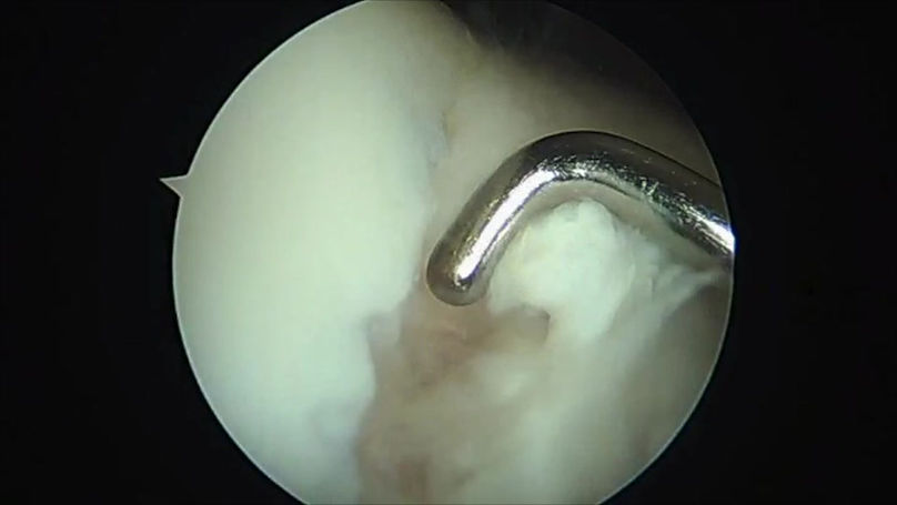
Osteochondral repair is used to treat a condition called Osteochondritis Dissecans (OCD). OCD is essentially a stress fracture that involves the bone and cartilage on the end of the femur (thigh bone). It usually begins in the early teen years and if it hasn't healed by the age of 15 it probably never will and the affected area will detach from the surrounding bone. If the affected piece is small it can simply be removed but if large needs to be repaired. Numerous techniques are used to reattach the loose piece depending on its size and how much bone is involved. Usually the detached piece is a big chunk of normal cartilage with a tiny sliver of dead bone on its undersurface. Getting this to heal requires biologic stimulus not just stability so bone grafting is usually required as well as reattaching the fragment. Biologically it behaves like a fracture so needs 6 weeks of non-weight bearing afterwards.
Large osteochondral fragment with a reciprocal defect in the end of the femur, fixed in place with sutures and osteochondral dowel grafts

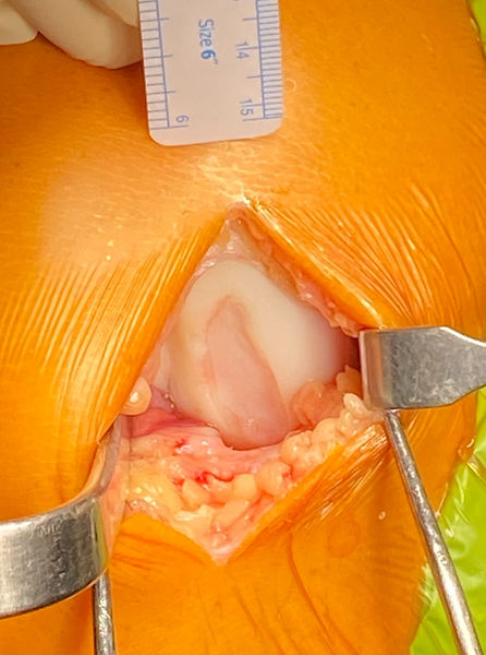
Autologous Osteochondral Transfer (OATS)
OATS involves taking dowels of cartilage and bone from a non-load bearing area of the knee and transplanting them into a cartilage defect. It is the only form of cartilage repair that guarantees normal articular cartilage attached to the underlying bone is in the defect. The bony portion of the transplant heals rapidly so after 4 weeks weight-bearing can begin and after 2 months no specific restriction are inflicted. The size of the lesion is limited to the amount of graft that can be taken. Three 1cm plugs is about the limit. OATS is also a good salvage option when other techniques have failed.
